



ESR1 mutations…
…are acquired
During endocrine treatment, breast cancer can evolve, resulting in the development of new mutations.1-4

…are subclonal
Molecular characteristics can vary between and within tumour sites. This is known as heterogeneity…5
…are different thanto intrinsic mutations
…as ESR1 mutations do not typically occur early in the course of the disease.3,6,7
…are acquired
During endocrine treatment, breast cancer can evolve, resulting in the development of new mutations.1-4
…are subclonal
Molecular characteristics can vary between and within tumor sites. This is known as heterogeneity…5
…are different thanto intrinsic mutations
…and may occur early in the course of the disease.3,6,7
What are ESR1 mutations?
ESR1 mutations are changes in the genetic code of the ESR1 gene that encodes the oestrogen receptor protein alpha (ERα).8
These mutations lead to an alteration in the structure of ERα that can affect how oestrogen binds to the receptor.5,8
ESR1 mutations can cause the ERα to be activated without having oestrogen bound to it, resulting in enhanced cancer growth, increased likelihood of metastasis, and the development of resistance to endocrine treatment (ET).5,8
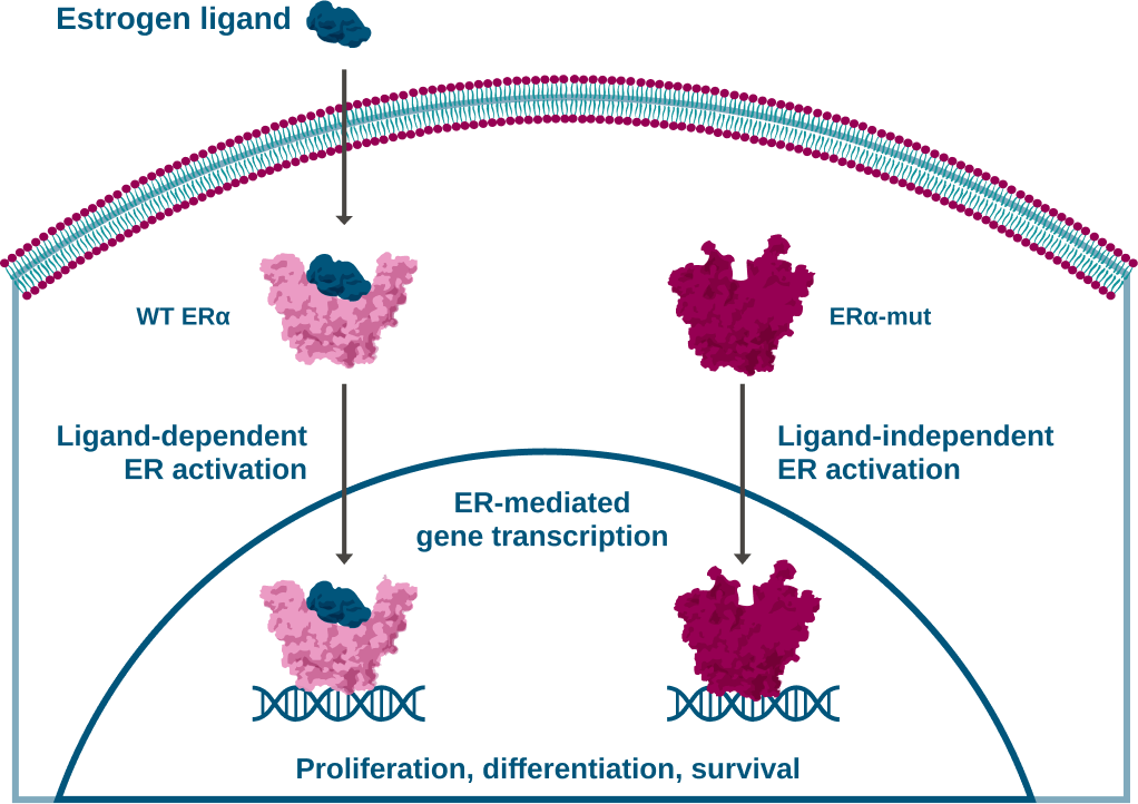
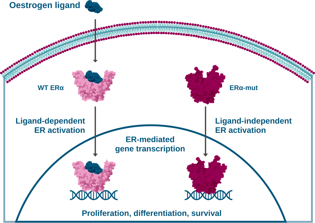

ESR1 mutations are acquired mutations
ESR1 mutations are driven by selective pressure resulting from the exposure to ET,1-4 and occur almost exclusively after aromatase inhibitor (AI) treatment in the metastatic setting.5,8-15


Longer exposure to ET increases the chance of acquiring ESR1 mutations in mBC.8,11-19
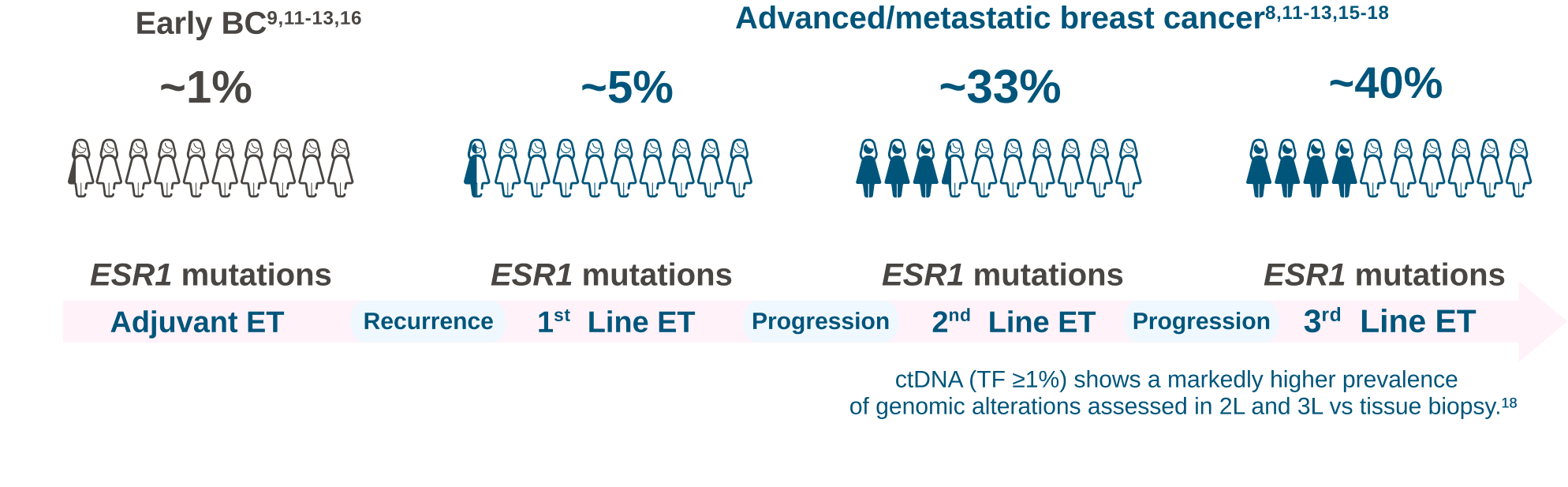
ESR1 mutations are rarely detected in early breast cancer or in a/mBC before 1L treatment.8-15
ESR1 mutations are associated with acquired resistance to AI treatment in the metastatic setting.8-15
ESR1 mutations are subclonal within the a/mBC tumour
a/mBC is a heterogenous disease and typically shows a high degree of intra-tumoural heterogeneity.5,8
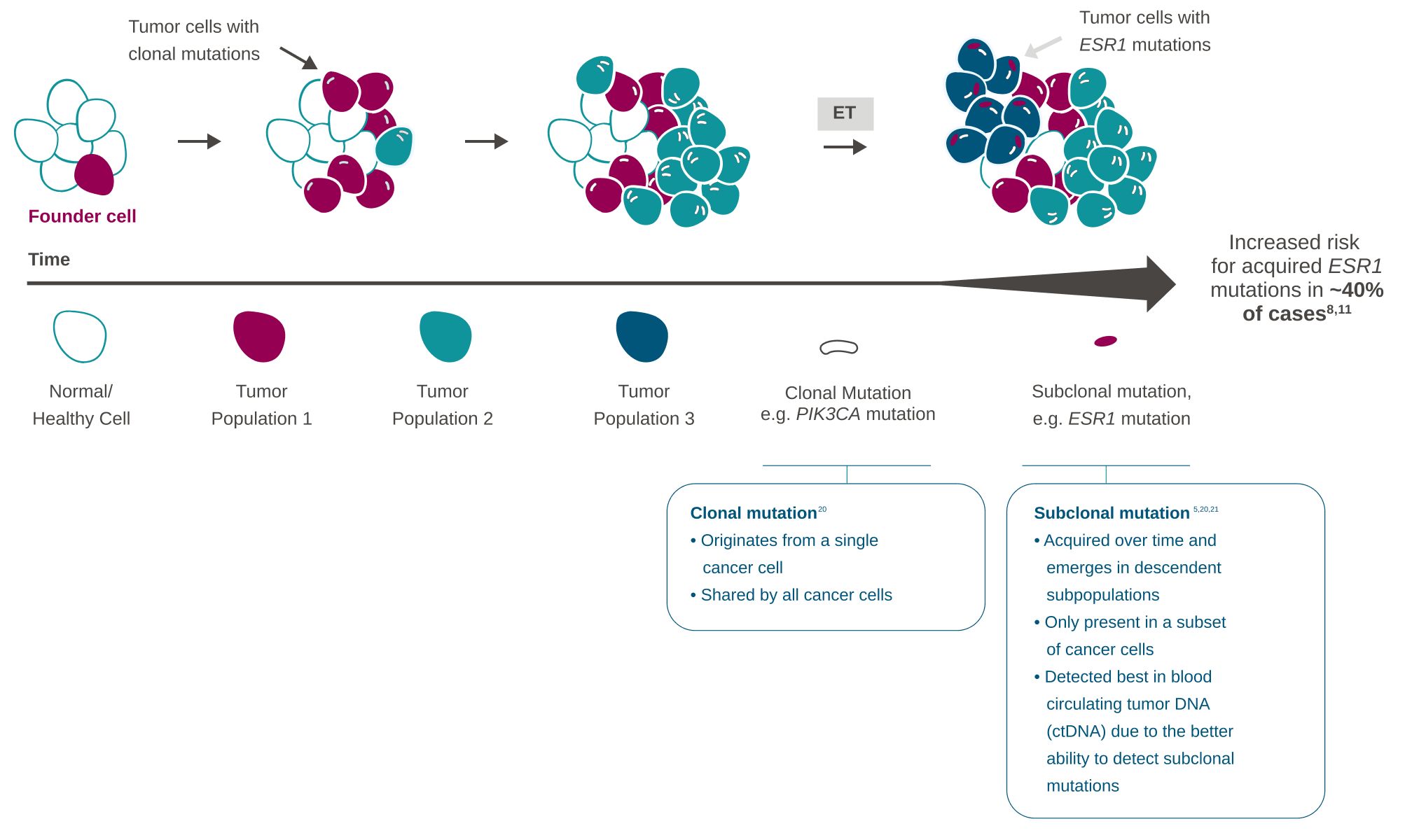
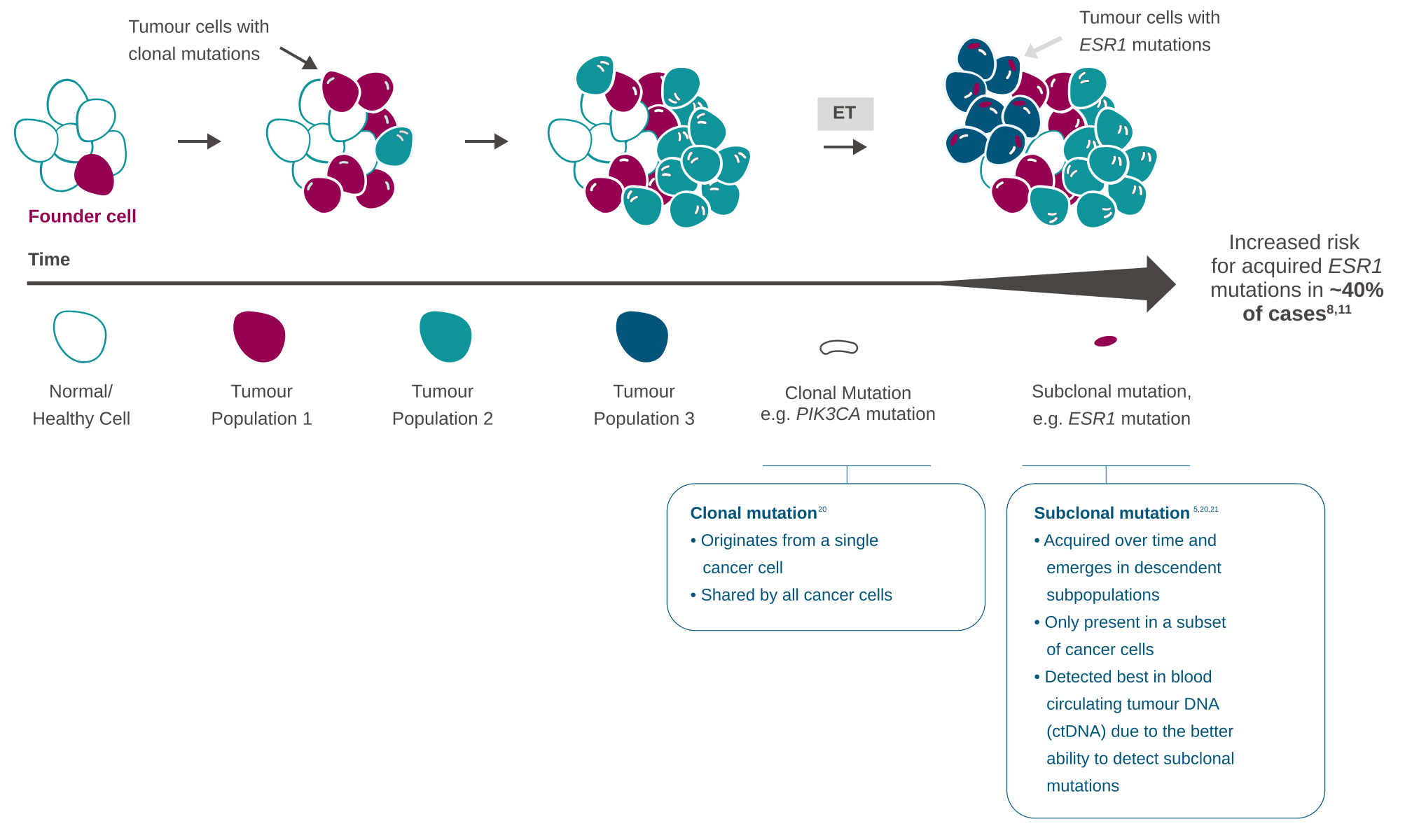
Subclones evolve and mutate over the course of the disease, resulting in tumour heterogeneity.21
ESR1 mutations are frequently subclonal in a/mBC.5
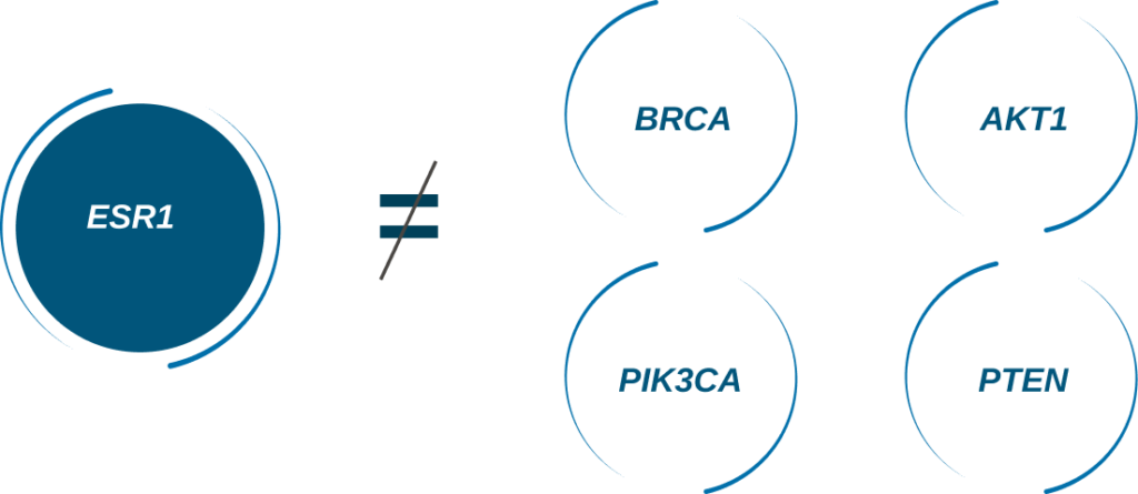
ESR1 mutations develop differently to other clinically relevant mutations in breast cancer
ESR1 mutations have a negative prognostic and predictive value, emerge during progression under endocrine treatment, and can be detected in the blood of patients.8,22 They are rarely found in primary tumours.9-12
Mutations such as AKT1, PIK3CA, and PTEN are often present at diagnosis therefore occurring before the influence of prescribed treatments.3,6,7
ESR1 mutations are subclonal (occur in a subset of cells) and heterogenous within the tumour, therefore it is possible that ESR1 mutations may not be detected in tissue biopsies.5,22
Roles of ESR1 mutations in patients with ER+/HER2- a/mBC23
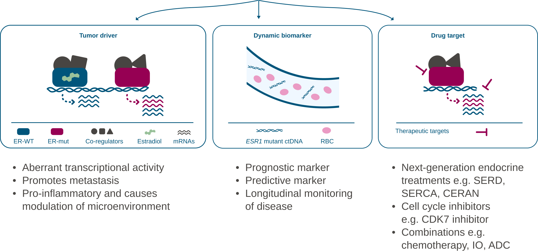
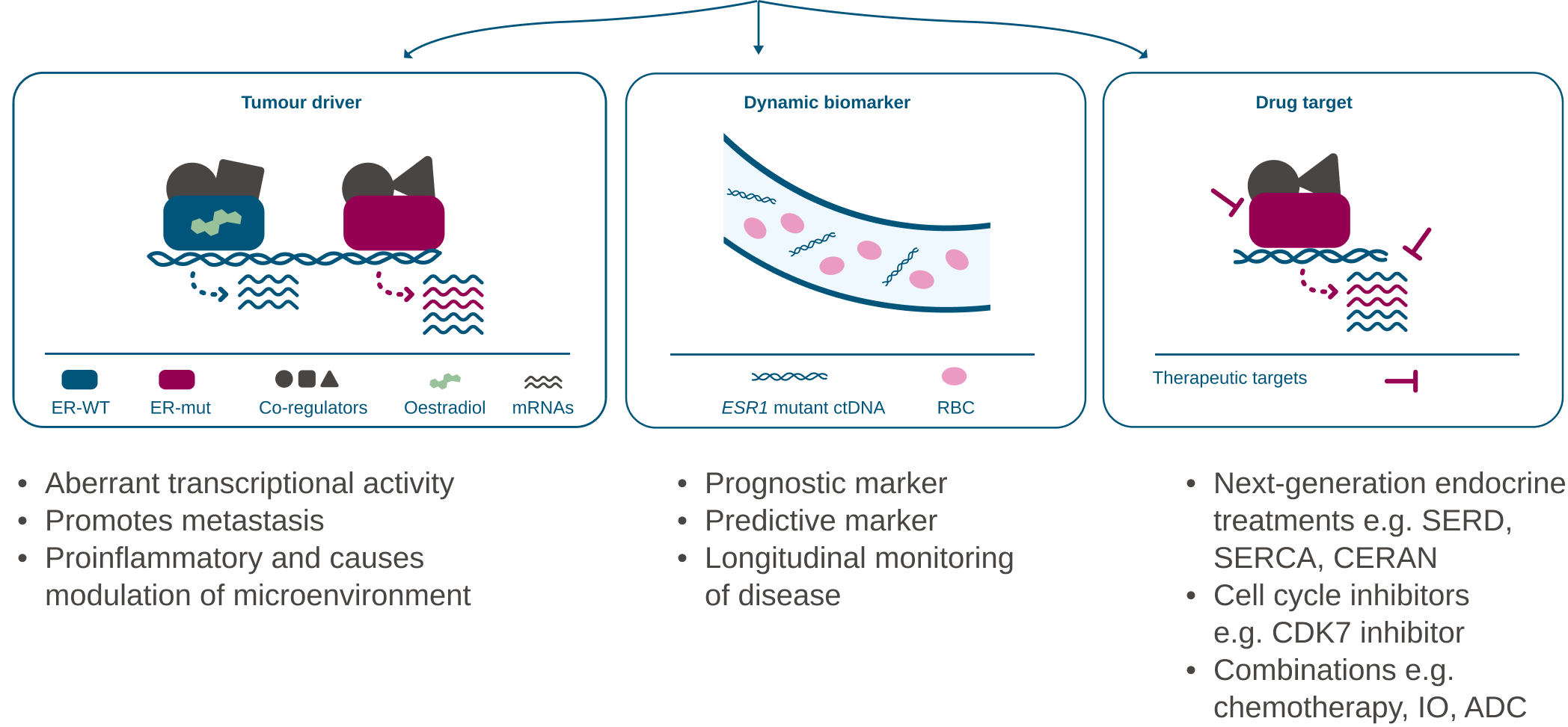
Adapted from Grinshpun et al. 2023.23
Common activating ESR1 mutations
ER+ breast cancer cells that have spread beyond the breast to metastatic sites have been found to either express wild-type ESR1 or harbour a variety of ESR1 alterations: ESR1 point mutations (most common), ESR1 amplifications and ESR1 fusions.24
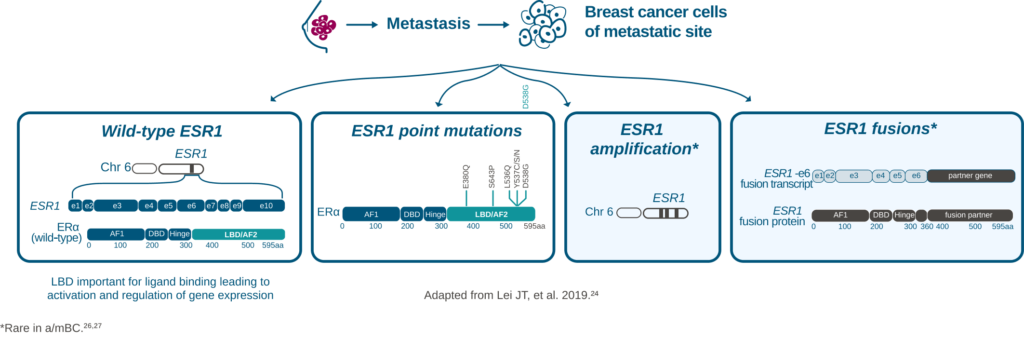
ESR1 point mutations
The most common genomic mechanism of acquired resistance to conventional endocrine treatment are activating missense mutations in ESR1.23
Activating mutations can lead to constitutive/ligand-independent transcriptional activities.23
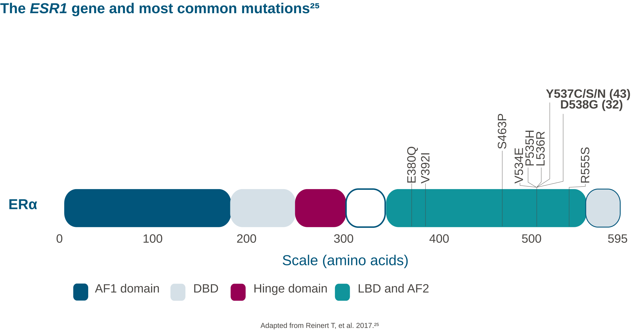
Most mutations that are pathogenic and putatively result in ET resistance are found in codons 536-538 of the ligand binding domain (LBD). The LBD is required not only for oestrogenic ligands but is also the domain that controls responses to anti-oestrogen antagonists.23
Y537S, D538G and Y537N account for 60–90% of the ESR1 mutations found in resistant cases. Other notable actionable mutations are E380Q and L536H, reported at a lower frequency.28
<i>ESR1</i> gene
The ESR1 (oestrogen receptor 1) gene is a gene located on chromosome 6 that codes for a protein called oestrogen receptor protein alpha (ERα).23
ERα binds to oestrogen and translocates to the nucleus to produce genomic actions.29
If these oestrogen-responsive genes are excessively activated, it could lead to the development of tumours and disease progression.23
ERα is widely expressed throughout the body,30,31 and plays a role in the pathophysiology of many cancers, including breast cancer.31
Human chromosome 6
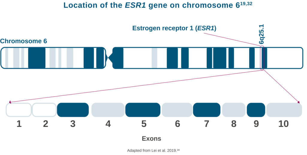
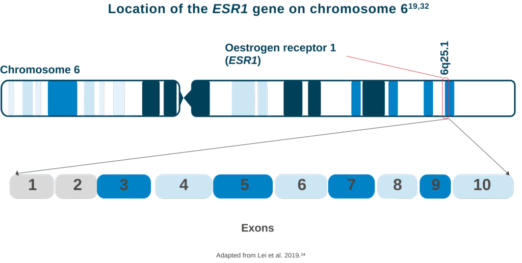
The ESR1 gene is located on chromosome 6, specifically at 6q25.19
The 8 coding exons 3-10 encode the transcription factor oestrogen receptor protein ERα.25,32
1L: first-line; ADC: antibody-drug conjugate; AF1/2: Activation Function 1/2; AI: aromatase inhibitor; AKT1: AKT Serine/Threonine Kinase 1; a/mBC: advanced or metastatic breast cancer; BC: breast cancer; BRCA: BReast CAncer gene; CD7: Cluster of Differentiation 7; CERAN: Complete OeEstrogen Receptor Antagonist; ctDNA: circulating tumour deoxyribonucleic acid; DBD: DNA binding domain; ER+/HER2-: oestrogen receptor-positive/human epidermal growth factor receptor 2-negative; ERα: oestrogen receptor alpha; ESR1: oestrogen receptor 1; ET: endocrine therapy; IO: immuno oncology; LBD: Ligand Binding Domain; mBC: metastatic breast cancer; mRNA: messenger ribonucleic acid; mut: mutation/mutated; PIK3CA: Phosphatidylinositol-4,5-Bisphosphate 3-Kinase Catalytic Subunit Alpha; PTEN: Phosphatase and TENsin homolog; RBC: red blood cell; SERCA: Sarco/Endoplasmic Reticulum Ca²⁺-ATPase; SERD: Selective OeEstrogen Receptor Degrader; TF: tumour fraction; WT: wild type.
- Clatot F, et al. Breast Cancer Res. 2020;22(1):56.
- Chandarlapaty S, et al. JAMA Oncol. 2016;2(10):1310.
- Turner NC, et al. Clinical Cancer Res. 2020;26(19):5172–7.
- Zundelevich A, et al. Breast Cancer Res. 2020;22(1):16.
- Dustin D, et al. Cancer. 2019;125(21):3714–28.
- Arthur LM, et al. Breast Cancer Res Treat. 2014;147(1):211–9.
- Turner NC, et al. N Engl J Med. 2023;388(22):2058–70.
- Brett JO, et al. Breast Cancer Res. 2021;23(1):85.
- Jeselsohn R, et al. Clinical Cancer Res. 2014;20(7):1757–67.
- Jeselsohn R , et al. Cancer Cell. 2018;33(2):173–86.e5.
- Allouchery V, et al. Breast Cancer Res. 2018;20(1):40.
- Schiavon G, et al. Sci Transl Med. 2015;7(313):313ra182.
- Toy W, et al. Nat Genet. 2013;45(12):1439–45.
- Jeselsohn R, et al. Nat Rev Clin Oncol. 2015;7(313):313ra182.
- Bidard FC, et al. J Clin Oncol. 2022;40(28):3246–56.
- Jhaveri K, et al. Ann Oncol. 2023;34(suppl_2):S338–S339.
- Lin N, et al. Ann Oncol. 2023;34(suppl_2):S338.
- Bhave MA, et al. Breast Cancer Res Treat. 2024;207(3):599–609.
- Lee N, et al. Int J Mol Sci. 2020;21(22):8807.
- Tarabichi M, et al. Nat Methods. 2021;18(2):144–55.
- El-Sayes N, et al. Cancers (Basel). 2021;13(4):806.
- Russano M, et al. J Exp Clin Cancer Res. 2020;39(1):95.
- Grinshpun A, et al. Biochem Biophys Acta Rev Cancer. 2023;1878(1):188830.
- Lei JT, et al. J Cancer Metastasis Treat. 2019;5:38.
- Reinert T, et al. Front Oncol. 2017;7:26.
- Heeke AL, et al. Breast Cancer Res Treat. 2022;196(2):279–89.
- Moelans CB, et al. Cell Oncol (Dordr). 2011;34(5):489–94.
- Silveira Vibert M, et al. Ann Oncol. 2023;34(S2):S383.
- Daniel JM, et al. Front Neuroendocrinol. 2023;0:101068.
- Sato K, et al. Front Endocrinol (Lausanne). 2023;14:1220150.
- Thomas C, et al. Nat Rev Cancer. 2011;11(8):597–608.
- Piscuolglio S, et al. Ann Oncol. 2018;29(4):787–9.